Gum disease is the most common health condition diagnosed in dogs, and one of the most consistently missed. By the age of three, the majority of dogs already show some degree of it. Yet because the early stages produce no obvious pain and dogs continue eating normally well into the advanced stages, most owners have no idea anything is wrong until the disease has progressed considerably.
Knowing what gum disease looks like in dogs is genuinely useful precisely because the condition announces itself visually before it announces itself behaviorally. The gums change color, texture, and shape in ways that are recognizable to anyone who knows what they are looking at, and those changes appear well before a dog starts dropping food or pawing at their face.
This guide covers what healthy gums look like as a baseline, how the appearance changes at each stage of disease, what the visible signs indicate about what is happening below the gumline, and what prevention and treatment involve.
Table Of Contents
- 1 What Healthy Gums Look Like
- 2 What Does Gum Disease Look Like in Dogs
- 3 How Gum Disease Progresses
- 4 Why It Matters Beyond the Mouth
- 5 Which Dogs Are Most at Risk
- 6 Dog Dental Care at Home
- 7 Professional Assessment and Treatment
- 8 When to Seek Attention
- 9 Checking Your Dog’s Mouth This Week
- 10 Frequently Asked Questions
What Healthy Gums Look Like
Establishing a baseline makes the abnormal far easier to spot.
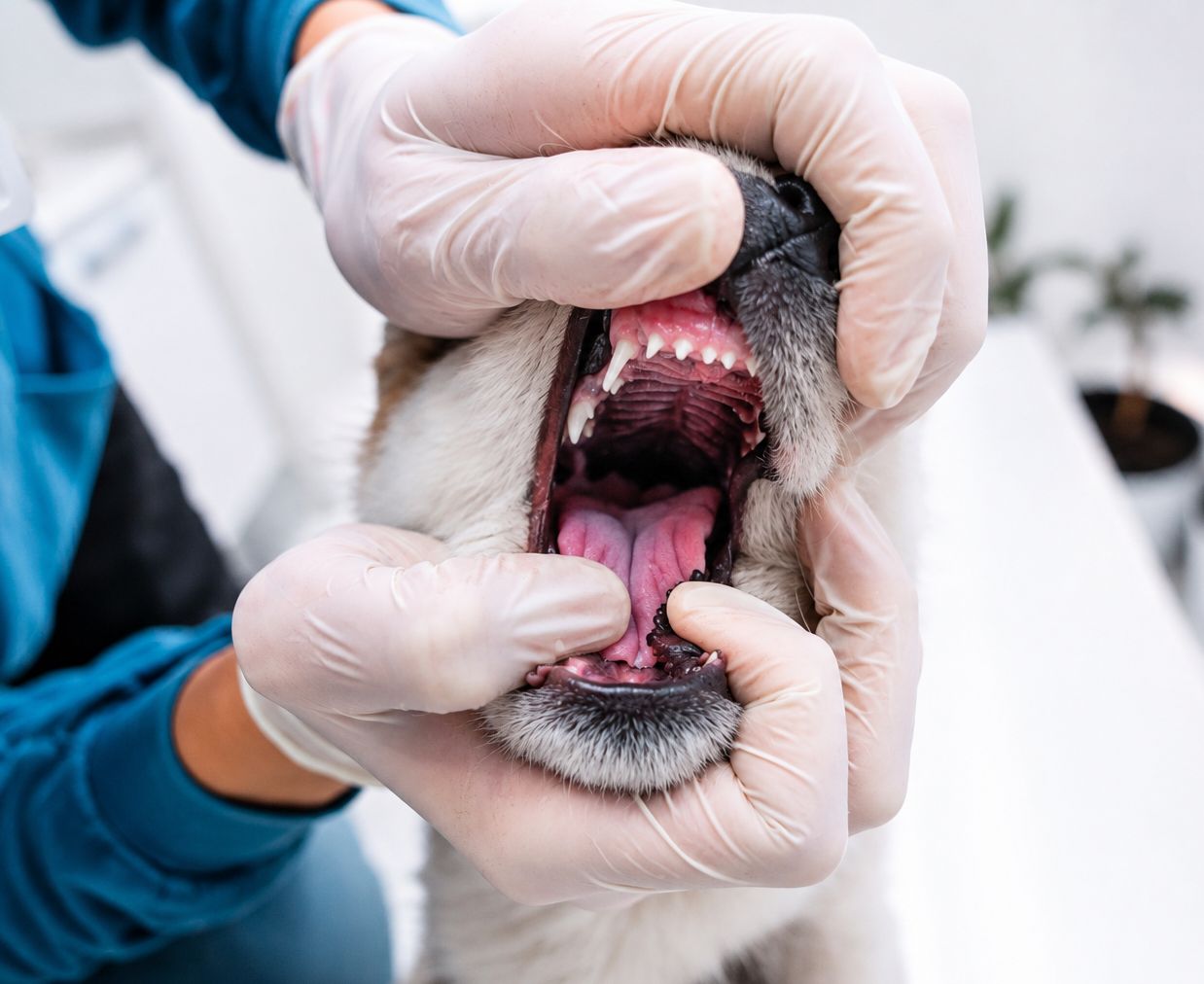
Healthy canine gums are pale pink, though some dogs naturally have areas of black or dark pigmentation which is entirely normal and unrelated to disease. The tissue is firm rather than puffy, and it meets each tooth in a clean, tapered edge that hugs the tooth surface closely.
The gum line should follow a smooth, even scalloped contour along the row of teeth. There should be no bleeding when the gums are touched gently, no visible swelling, and no gaps between gum and tooth. Teeth themselves should be white to off-white without visible buildup at the base.
Lifting your dog’s lip once a week takes seconds and builds familiarity with what normal looks like for your particular dog, which is what makes changes noticeable.
What Does Gum Disease Look Like in Dogs
Redness Along the Gum Line
The earliest visible change is a thin band of redness where the gums meet the teeth. Rather than the uniform pale pink of healthy tissue, a distinct red line appears along the margin, often most noticeable on the outer surfaces of the upper back teeth where plaque accumulates fastest.
This is gingivitis, inflammation of the gum tissue caused by bacterial plaque sitting along the gumline. At this stage the condition is confined to the soft tissue and is fully reversible with appropriate treatment. This is the single most important window for intervention.
Swollen or Puffy Gum Tissue
As inflammation progresses, the gum tissue loses its firm, tapered edge and becomes visibly swollen. Instead of hugging the tooth closely, the margin appears rounded, thickened, and slightly raised. The clean scalloped contour becomes irregular.
Swelling indicates the inflammatory response has intensified and the tissue is becoming compromised rather than simply irritated.
Bleeding
Healthy gums do not bleed from gentle contact. Gums that bleed when touched, when a toy is chewed, or that leave pink traces on food or water bowls indicate that the tissue has become fragile through sustained inflammation.
Some owners notice this first as small pink smears on a chew toy or a rope. It is an unambiguous sign that gum disease is established rather than beginning.
Visible Tartar Buildup
Plaque is a soft, colorless film that forms continuously on tooth surfaces. When it is not removed, minerals in the saliva harden it into tartar, also called calculus, which is visible as yellow, tan, or brown deposits at the base of the teeth.
Tartar matters for two reasons. Its rough surface gives new plaque an ideal texture to adhere to, accelerating accumulation. And it cannot be brushed away once formed, requiring professional removal. What is visible above the gumline also indicates that similar deposits have formed below it, where the real damage occurs.
Receding Gums and Exposed Roots
As periodontal disease in dogs advances beyond the soft tissue, it begins destroying the structures anchoring the teeth. Visually this appears as the gum line pulling back from the tooth, exposing progressively more of the tooth surface and eventually the root.
Teeth may look noticeably longer than neighboring ones. In severe cases the root furcation, the point where the roots of a multi-rooted tooth divide, becomes visible as a gap at the base of the tooth. This indicates substantial bone loss and is not reversible.
Pus and Discharge
Visible pus at the gum margin, or discharge when the gum is gently pressed, indicates active infection within the periodontal pocket. Pockets form as the attachment between gum and tooth breaks down, creating spaces that trap bacteria and debris beyond the reach of brushing.
Loose or Missing Teeth
In an adult dog, a tooth that moves when touched is not normal. Mobility indicates that the bone and ligament holding it in place have been substantially destroyed. Teeth at this stage are painful and typically require extraction.
The Smell
Persistent bad breath is often the sign that prompts an owner to look more closely. A mild food-related odor after eating is normal. A consistently foul or unusually strong smell is produced by the bacteria driving periodontal disease and the tissue breakdown accompanying it.
Breath odor is worth taking seriously rather than accepting as a normal feature of dog ownership. It is frequently the first indication owners get that something below the gumline needs attention.
How Gum Disease Progresses
Periodontal disease is graded in four stages, each with a characteristic appearance and a different outlook.
Stage |
What You Can See |
What Is Happening Below |
Reversible? |
| Stage 1: Gingivitis | Thin red line along gum margin, mild swelling | Inflammation confined to gum tissue, no bone loss | Yes, fully |
| Stage 2: Early periodontitis | Redness, swelling, bleeding, visible tartar | Early attachment loss, up to a quarter of support lost | Damage is not reversible, progression can be halted |
| Stage 3: Moderate periodontitis | Gum recession, larger tartar deposits, possible discharge | Significant bone loss, pocket formation around roots | No, treatment aims to preserve remaining structure |
| Stage 4: Advanced periodontitis | Marked recession, exposed roots, loose teeth, pus | Severe bone destruction, teeth losing anchorage | No, extraction usually required |
The critical distinction sits between stage one and stage two. Gingivitis involves only soft tissue and resolves completely with professional cleaning and consistent home care. Once the disease crosses into periodontitis, the bone and ligament that have been lost do not regenerate. Treatment from that point aims to stop further destruction rather than to restore what is gone.
Why It Matters Beyond the Mouth
Periodontal disease is not confined to the mouth. The inflamed, ulcerated tissue within periodontal pockets provides a direct route for oral bacteria to enter the bloodstream, and research in veterinary medicine has identified associations between periodontal disease and changes affecting the heart, kidneys, and liver.
There is also the matter of pain. Dogs are remarkably stoic about oral discomfort and will continue eating through significant dental pain, which is precisely why owners so often report that their dog “was fine” right up until a dental procedure. Many report afterward that their dog seems brighter, more playful, and more interested in food than they had been for months, revealing discomfort that had been present but invisible.
Because dental disease affects and is affected by general health, assessment forms part of routine wellness examinations rather than being treated as a separate concern.
Which Dogs Are Most at Risk
Some dogs accumulate dental disease considerably faster than others.
- Small and toy breeds face substantially elevated risk, as their teeth are often crowded within a smaller jaw, creating tight spaces that trap plaque and are difficult to clean.
- Brachycephalic breeds with shortened muzzles frequently have rotated or overlapping teeth, producing the same crowding problem and accelerating accumulation.
- Senior dogs have simply had more years for plaque and tartar to build, and often have earlier-stage disease that has been progressing quietly for a long time.
- Dogs fed exclusively soft food miss the mild mechanical cleaning that chewing provides, though it is worth noting that no diet substitutes for brushing.
- Dogs with no established home dental routine accumulate tartar considerably faster, as plaque hardens within days when it is not disrupted.
Dog Dental Care at Home
Brushing
Daily brushing with a pet-formulated toothpaste is the single most effective thing an owner can do. Human toothpaste must not be used, as it contains ingredients including xylitol and fluoride that are harmful if swallowed.
Introducing brushing gradually produces far better long-term compliance than attempting a full session immediately. Letting the dog taste the toothpaste, then touching the brush briefly to a few teeth, then building up over a week or two, establishes it as an unremarkable routine rather than a struggle. The outer surfaces of the upper back teeth deserve the most attention, as this is where tartar accumulates fastest.
Frequency matters because plaque begins hardening into tartar within roughly two to three days. Brushing weekly does not prevent tartar formation. Daily brushing does.
Other Supportive Measures
Veterinary-approved dental chews provide mechanical abrasion that helps reduce plaque, and dogs generally enjoy them. Water additives and oral rinses formulated for dogs offer additional antibacterial support and are useful for dogs who resist brushing entirely.
These measures support brushing rather than replacing it. A dog receiving only chews will accumulate disease more slowly than one receiving nothing, but considerably faster than one being brushed daily.
Worth avoiding are excessively hard chew objects including antlers, bones, and hard nylon toys, which are a common cause of fractured teeth. A useful guideline is that if you cannot indent it with a thumbnail, it is hard enough to break a tooth.
Professional Assessment and Treatment

What a Dental Examination Covers
A veterinarian examining your dog’s mouth assesses the gum tissue for inflammation, recession, and pocket formation, checks each tooth for mobility, discoloration, and fractures, evaluates tartar accumulation, and looks for oral masses or ulceration.
This assessment identifies the stage of disease present and determines what treatment is needed. Because it does not require anesthesia, it can be performed comfortably during a routine home visit, which is particularly valuable for dogs who find clinic environments stressful. The broader advantages of assessing dogs in their own environment are covered in the guide on benefits of at-home vet services for anxious pets and busy owners.
Comfort Paws provides pet dental assessments that identify disease, gingivitis, and tooth decay, with treatment recommendations and referral coordination for procedures requiring specialist facilities.
What Requires a Clinic
Professional cleaning to remove tartar from below the gumline requires general anesthesia. This is not a matter of convenience. The area that matters most in periodontal disease is beneath the gum margin, and cleaning it thoroughly requires a still patient and a protected airway.
Dental radiographs, which reveal bone loss and root disease invisible on external examination, similarly require anesthesia. So do extractions.
A note on anesthesia-free dental cleaning, which is sometimes marketed as a gentler alternative: it removes visible tartar from the crown while leaving the subgingival deposits that cause periodontal disease entirely untouched. The result looks improved while the disease continues progressing beneath the gumline. Veterinary dental organizations advise against it for this reason.
When to Seek Attention
Certain findings warrant prompt evaluation rather than waiting for a routine appointment:
- Facial swelling, particularly beneath the eye or along the jaw, which commonly indicates a tooth root abscess requiring timely treatment.
- Refusing food for more than a day, as oral pain severe enough to prevent eating needs assessment and pain management.
- Visible pus or blood in the mouth that does not resolve, which indicates active infection or a lesion requiring examination.
- Teeth that are visibly loose or have shifted position, indicating substantial periodontal destruction.
- Pawing at the face or marked sensitivity when the muzzle is touched, which reflects pain the dog is otherwise concealing.
For less urgent findings including persistent bad breath, visible tartar, or gum redness, scheduling a dental assessment at your next available appointment is appropriate. Dogs who are unwell can be seen through a sick visit.
Checking Your Dog’s Mouth This Week
Gum disease is one of the few common conditions in dogs where an owner can see the earliest stage with their own eyes, at home, in under a minute. That makes it unusually preventable compared with the internal conditions that require testing to detect.
The stage that matters is the first one. A thin red line along the gum margin is entirely reversible. Left alone, it becomes bone loss that is not. The difference between those two outcomes is often nothing more than whether someone looked.
If you have noticed changes in your dog’s gums, persistent bad breath, or visible tartar, Comfort Paws Veterinary Care provides at-home dental assessments in Brooklyn with clear guidance on what is present and what to do about it. Reach out to schedule a consultation to have your dog’s oral health assessed without the stress of a clinic visit.
Frequently Asked Questions
Q: What does gum disease look like in dogs at the earliest stage?
A: The earliest visible sign is a thin red line where the gums meet the teeth, often with mild swelling along the margin. This is gingivitis, and it is fully reversible with professional cleaning and consistent home care.
Q: Can gum disease in dogs be reversed?
A: Gingivitis, the first stage, resolves completely with treatment. Once periodontal disease in dogs progresses to bone loss, that damage is permanent. Treatment then focuses on halting further destruction rather than restoring what has been lost.
Q: How often should I brush my dog’s teeth?
A: Daily brushing is the standard recommendation, because plaque begins hardening into tartar within two to three days. Less frequent brushing slows accumulation but does not prevent tartar forming, which then requires professional removal.
Q: Is bad breath in dogs normal?
A: A mild food-related odor after eating is normal. Persistent foul breath is not, and usually indicates bacteria associated with periodontal disease. It is one of the most common early signs that prompts owners to seek a dental assessment.
Q: Can a dog’s teeth be cleaned without anesthesia?
A: Anesthesia-free cleaning removes visible tartar from the crown but leaves deposits below the gumline where periodontal disease actually occurs. The mouth looks better while disease progresses underneath, which is why veterinary dental organizations advise against it.